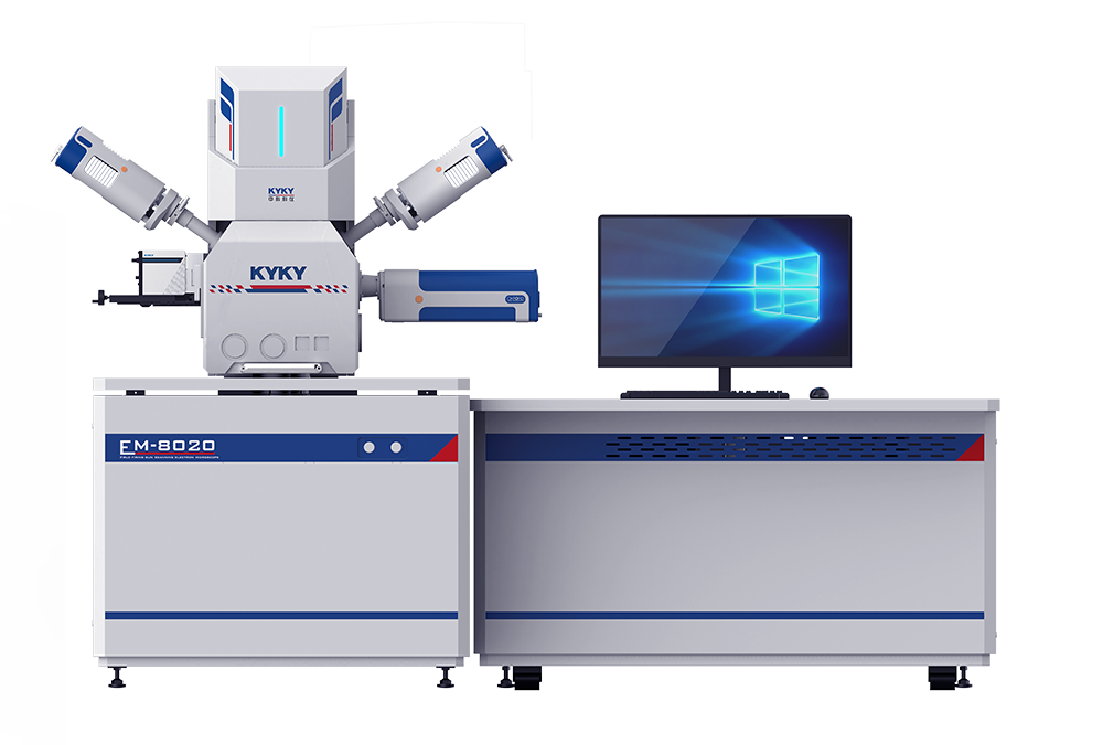
Cell and tissue observation
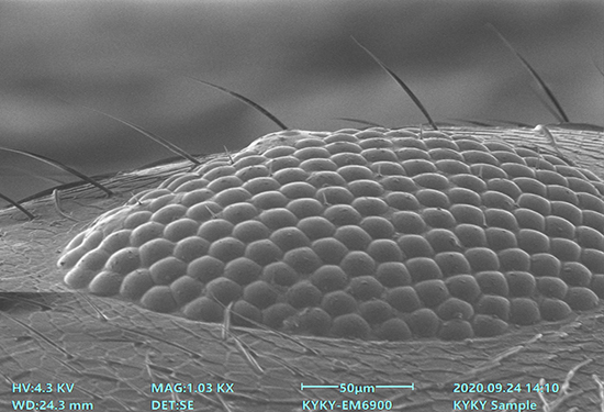
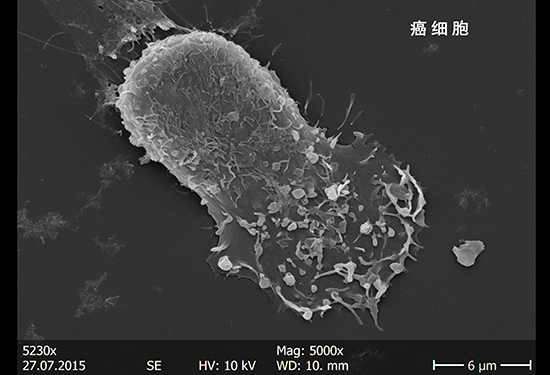
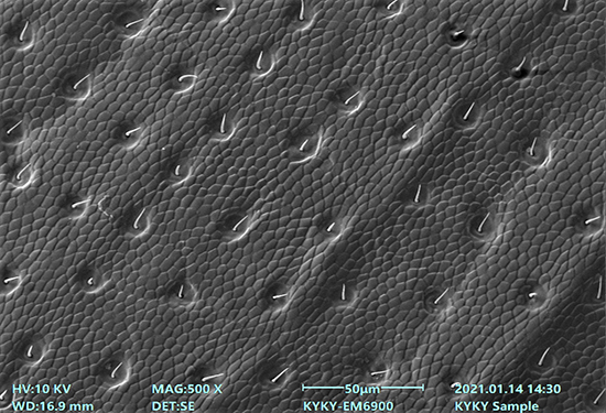
◆Cellular morphology and structure analysis: The scanning electron microscope can be used to observe the surface morphology of cells, including the morphology and position distribution of cell membranes and organelles in the cytoplasm (e.g., mitochondria, chloroplasts, etc.) at very high resolution. It is of great significance for understanding the physiological functions and pathological changes of cells.
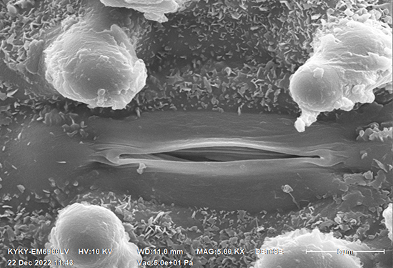
◆Tissue structure and function studies: The scanning electron microscope can be used to observe the microstructure of different tissues such as epithelial tissue, connective tissue, etc., and how these structures support the function of the tissues. For example, in the medical field, it can be used to study bone structure, blood vessel wall structure, etc.
Microbiological studies
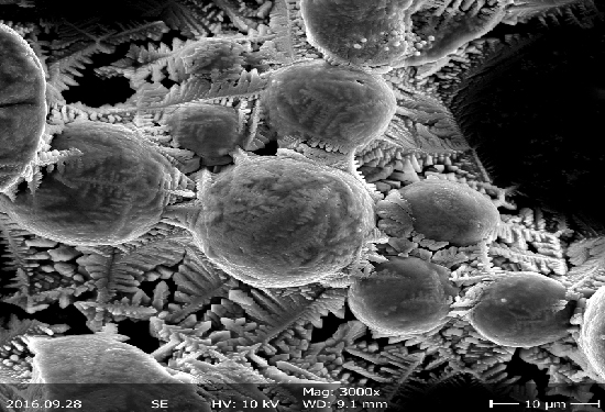
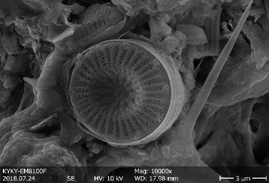
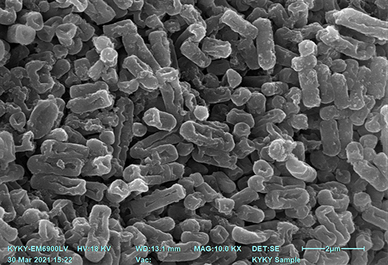
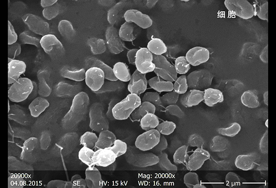
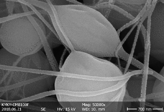
◆Microbial morphology observation: The scanning electron microscope can be used to observe the morphology of bacteria, fungi and other microorganisms, including the observation of their cell wall structure, flagella, cilia and other microstructures. It is of great value for the classification and identification of microorganisms and ecological studies.
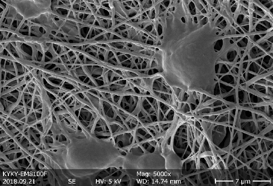
◆Microorganism-host interactions: The scanning electron microscope can be used to observe the attachment, invasion and reproduction process of microorganisms on host cells or tissues, and to reveal the interaction mechanism between microorganisms and hosts.
Biomedical applications
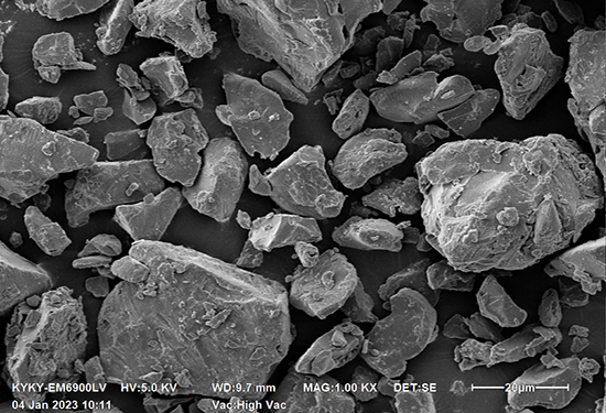
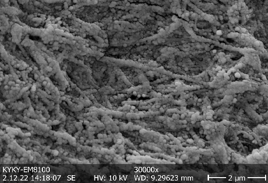
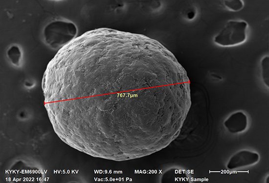
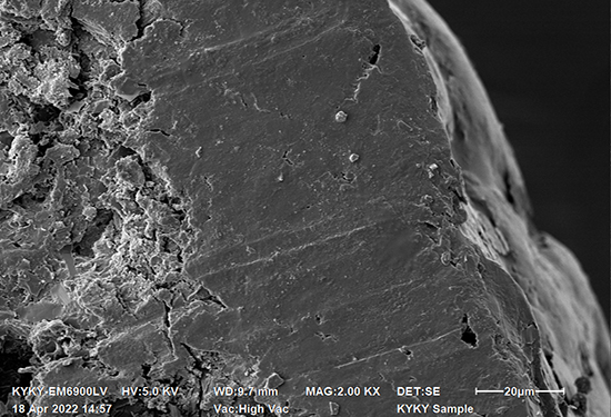
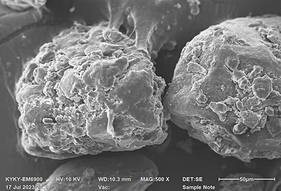
◆Biomaterials studies: The scanning electron microscope can be used to study the surface morphology, pore structure and other properties of biomaterials to optimize the performance and application effect of biomaterials.
◆Disease diagnosis: The scanning electron microscope has become an indispensable scientific research tool and means in the study of medical morphology. It can be used for electron microscope diagnosis of difficult and complicated diseases, such as assisting doctors in diagnosis by observing the microstructure of pathological tissues.
◆Research and development of drugs: In the research and development of drugs, the scanning electron microscope can be used to observe the effects of drugs on cells or tissues, including drug absorption, distribution, metabolism, and excretion, providing important information for drug design and optimization.
◆Tissue engineering and regenerative medicine: The scanning electron microscope can be used to observe the microstructure of tissue engineering products, such as artificial skin, artificial blood vessels, etc., to evaluate their quality and function. Meanwhile, it can also be used to study the morphologic changes during stem cell differentiation.